|

Home > Research


Research Overview


 |
The drive to break down and analyze objects in their most basic forms is one of the defining characteristics of our innate curiosity and desire to understand the world on the most fundamental level. Tremendous societal benefits can be gained by satisfying this curiosity. For example, the ability to observe, detect and manipulate single biomolecules and macromolecules can be exploited for numerous purposes, including early disease detection and drug design to improve the quality and length of life. Microscopes have long helped curious humans look at the very small scale, but what if we could replace costly and bulky high-end microscopes with tiny chips that can detect, analyze, and manipulate single biomolecules? Such a miniaturization can transform both basic scientific research and the design and utility of biomedical devices. Research in the W.M. Keck Center for Nanoscale Optofluidics combines integrated optofluidics with advanced nano-fabrication to build such chips and use them in novel studies single biomolecules. Optofluidics is an emerging field of research that involves the interaction of light and fluids. Combining techniques from integrated optics and microfluidics allows for routing both optical signals and liquid samples in the plane of the chip. This provides a unique opportunity for developing a new class of devices for biology and biomedicine with unprecedented capabilities. In this Center, experts in integrated optofluidics, nanoparticle synthesis, nanopore technology, and molecular biology of RNA work together to create a novel class of devices relying entirely on integrated optical technology as opposed to bulk microscopy. For more information on ongoing projects, please follow the links to the individual research labs. |
| Research Highlights |
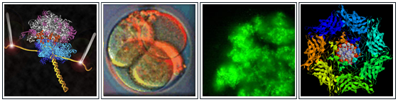
Single virus detection on optofluidic chips: M.I. Rudenko et al. demonstrate optical detection of single bacterial viruses on an optofluidic chip (Biosensors and Bioelectronics 24, 3258-3263 (2009)). Controlled enzyme binding to single DNA molecules: Researchers in the Akeson and Dunbar groups show that ionic current signatures in a hemolysin nanopore can be used to implement robust analysis of the assembly of complexes between DNA and RNA processing enzymes and their substrates at the single molecule level (ACS Nano 3, 995–1003 (2009)). Novel on-chip trap for optical particle analysis: The Schmidt group introduces a novel optical trap based on loss in liquid-core optical waveguides that allows for particle position control along an optofluidic channel and fluorescence studies on trapped particles (Lab on Chip 9, 2212 (2009)). Dynamics of RNA translation in single ribosomes: Ermolenko et al. measure details of the translation process in single ribosomes using optical fluorescence techniques (FRET). (Mol Cell. 30:578-88 (2008)). Hollow gold nanospheres show promise cancer detection: Researchers in the Zhang group collaborated with colleagues at the MD Anderson Cancer Center (Texas) to selectively bind functionalized hollow gold nanoshells to cancer cells in mice and kill the cells with a strong optical pulse at the nanoshell absorption frequency. (http://www.ucsc.edu/news_events/text.asp?pid=2790). Observation of translation in single ribosome: A collaboration between the Noller and Tinoco (UCB) groups reports the first observation of an unraveling RNA strand one codon at a time using optical tweezer technology. (Nature, 452:598-603 (2008)). |